4 min read
What Is a Functional Brain Map?

Picture your brain as a complex city—streets of thoughts, highways of memory, neighborhoods full of movement, language, and emotion. A functional brain map lets us see exactly where each of those functions lives in your brain.
A functional brain map is a clinical tool used to identify and visualize brain function in real time. It helps neurologists and neurosurgeons locate eloquent areas—the regions of the brain responsible for critical abilities like speech, memory, and motor control.
By mapping how your brain functions, doctors can plan safe, personalized treatments, whether for surgery, seizures, or recovery from trauma.
Key Concepts in Brain Mapping
Brain mapping isn’t just about anatomy—it’s about action.
This technique reveals the brain cerebral cortex function, showing how areas light up when you move a finger, recall a face, or speak a word. These insights shape everything from diagnosis to surgical precision.
Difference Between Functional and Structural Brain Maps
Let’s clear this up:
- A brain function map shows how parts of the brain work.
- A structural map shows what the brain looks like physically.
Think of it like a power grid: a structural map shows the wiring; a functional brain map shows where the electricity flows.
Why Functional Brain Maps Are Important
Would you trust a pilot who can’t see the runway?
Exactly.
That’s why brain mapping is essential before any surgery or treatment near sensitive brain regions. It tells doctors where not to go and how to protect you.
Applications in Neurosurgery and Brain Disorders
A functional brain map is commonly used to:
- Prepare for brain tumor or epilepsy surgeries
- Assess brain cerebral cortex function after injury
- Guide stroke rehabilitation
- Plan deep brain stimulation in Parkinson’s disease
Example: A patient with seizures was found to have abnormal brain activity near the motor cortex. Using brain mapping, surgeons avoided the area, controlling seizures without affecting movement.
Preserving Brain Function During Surgery
A functional brain map helps surgeons stay clear of areas linked to speech, movement, or memory—functions that define a person’s quality of life.
Your surgeon isn’t just operating on tissue—they’re protecting who you are.
Brain Mapping Techniques Used in Clinical Settings
Different methods help create your personal functional brain map.
Awake Craniotomy and Cortical Mapping
Yes, it’s possible to be awake during brain surgery—and it can save your life.
During awake craniotomy, patients perform simple tasks like speaking or moving while doctors stimulate brain regions. This allows real-time functional mapping and protects vital skills.
Imaging Tools and Coordinate Systems
Other brain mapping tools include:
- fMRI (functional MRI) – reveals activity by tracking blood flow.
- ECoG (electrocorticography) – detects electrical signals in the brain.
- Coordinate systems – align your brain function map with known anatomical landmarks.
These tools work together to build a full picture of your brain cerebral cortex function.

Functional Brain Mapping Tools
|
Tool |
Type |
Advantages |
Disadvantages |
Best Use Case |
|
fMRI |
Non-invasive |
Visualizes brain activity without surgery |
Expensive; slower updates |
Mapping memory, planning speech surgeries |
|
ECoG |
Semi-invasive |
High accuracy for electrical activity |
Requires craniotomy |
Seizure mapping, surgical planning |
|
MEG |
Non-invasive |
Good temporal resolution |
Less available, costly |
Functional brain timing studies |
|
Awake Craniotomy |
Invasive |
Direct mapping with patient feedback |
Complex procedure |
Tumor removal near brain function areas |
|
TMS |
Non-invasive |
Non-surgical, effective for motor mapping |
Lower spatial precision |
Quick assessment of brain mapping needs |
Areas Mapped in the Functional Brain Map
Let’s explore what areas are highlighted when creating a brain function map.
Language and Speech
Brain mapping can pinpoint language zones in the brain cerebral cortex, typically in the left hemisphere. Protecting these during surgery prevents loss of communication ability.
Sensory and Motor Control
These areas manage your touch, movement, and coordination. If your surgeon knows exactly where they are thanks to a functional brain map, they can avoid accidental damage.
Memory and Cognition
Want to protect your ability to remember names or events?
Mapping your brain function allows doctors to work around memory centers like the hippocampus, which can be damaged by tumors or trauma.
Challenges and Limitations
Even with advanced tools, brain mapping has its challenges.
Individual Brain Variation
No two people have the same brain function map. This is why personalization is key—generic maps won’t protect your unique brain.
Dealing with Brain Lesions and Injuries
Lesions and swelling can distort normal brain mapping. But with skilled neurologists, these challenges can be overcome with updated imaging and customized plans.
Functional Brain Mapping at Neurology Mobile, Miami
Looking for expert functional brain mapping in Miami?
We’ve got you covered.
At Neurology Mobile, we combine cutting-edge technology with personalized care to deliver the most accurate brain function maps available today.
Patient-Centered Brain Mapping Procedures
We tailor each procedure to your needs—whether you’re managing seizures or preparing for surgery. Our team uses non-invasive tools to map your brain cerebral cortex function with precision and care.
Importance of Custom Brain Maps for Treatment Planning
A customized functional brain map ensures your treatment is safe, effective, and aligned with your personal brain layout.
Because protecting your brain function is protecting your life.
Ready to Take the Next Step?
A functional brain map isn’t just a medical tool—it’s a window into who you are.
From preserving speech to improving treatment outcomes, brain mapping gives doctors the power to treat your condition without compromising your identity.
So ask yourself:
If your brain needed help, wouldn’t you want the clearest possible guide?
At Neurology Mobile, we offer personalized functional brain mapping services designed to protect and empower you.
📞 Call now or schedule your consultation to discover how our advanced brain function mapping can change your life.
Your brain deserves nothing less.
Frequently Asked Questions About Functional Brain Mapping
What is functional mapping of the brain?
Functional brain mapping is a technique used to identify which areas of the brain are responsible for critical functions like speech, movement, memory, and sensation. Unlike structural imaging, which shows the anatomy of the brain, functional mapping reveals how different areas of the brain work. This is especially useful in preparing for brain surgery, helping doctors avoid important regions and reduce the risk of complications. Techniques like fMRI, ECoG, and awake craniotomy are commonly used for this purpose.
What are the functional areas of the brain?
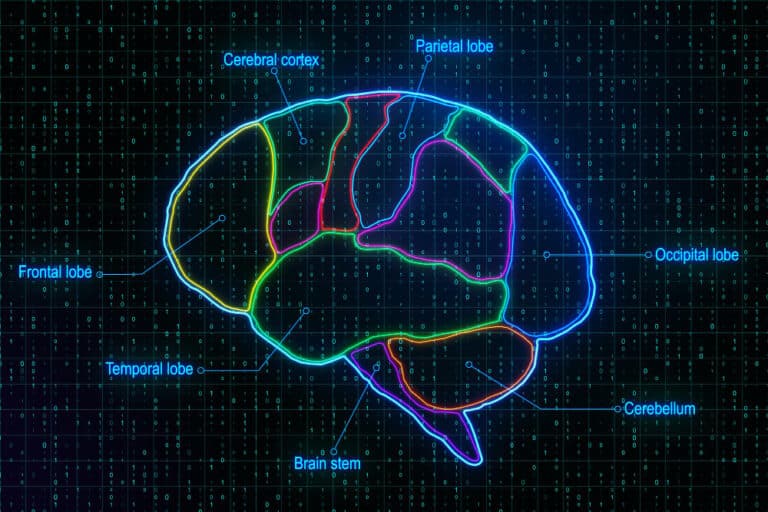
The brain is divided into several functional areas, each with its own job:
- Frontal lobe: responsible for decision-making, problem-solving, and motor control.
- Temporal lobe: involved in language and auditory processing.
- Parietal lobe: manages sensory perception and spatial awareness.
- Occipital lobe: handles visual information.
- Cerebellum: coordinates balance and movement. Functional brain mapping helps identify the precise locations of these functions in each individual, which is important because every brain is slightly different.
Does brain mapping really work?
Yes, brain mapping is a proven and reliable tool in modern neurology and neurosurgery. It helps doctors pinpoint vital areas of the brain to improve outcomes in surgeries and treatments for epilepsy, brain tumors, and other neurological conditions. Clinical studies and decades of practice have shown that functional mapping significantly reduces surgical risks and improves recovery times by preserving essential brain functions.
Is functional brain mapping painful?
No, most functional brain mapping techniques are non-invasive and painless. Procedures like fMRI and transcranial magnetic stimulation (TMS) don’t require surgery or discomfort. Even in cases where awake brain surgery is needed, patients are kept pain-free and comfortable using anesthesia. Your doctor will explain the process clearly and ensure you’re relaxed throughout the procedure.
Who should consider getting a functional brain map?
Functional brain mapping is recommended for people who:
- Are undergoing brain surgery (especially near language or motor areas)
- Have epilepsy that doesn’t respond to medication
- Are being treated for brain tumors or lesions
- Need precise planning for stroke rehabilitation It’s also valuable in research and personalized medicine to better understand cognitive function and neurological health.
