4 min read
Electrocorticography (ECoG): An In-Depth Guide
Electrocorticography, commonly known as ECoG, represents a pivotal development in the realm of neurophysiological monitoring. The origins of ECoG can be traced back to the early 20th century. While the first recordings of electrical activity from the human brain’s surface came from Hans Berger’s invention of the electroencephalogram (EEG) in the 1920s, the idea of directly placing electrodes on the exposed surface of the brain to capture electrical activity emerged several decades later.
This approach was initially used by neurosurgeons in the 1950s as a means to locate epileptic foci during surgical procedures. Over the years, as surgical techniques and electrode technology evolved, ECoG started to gain traction not only as a clinical tool but also as a valuable method for brain research. Its capacity to provide high-resolution spatial and temporal data made it an invaluable asset for understanding the intricacies of the human brain.
Basic principles of ECoG
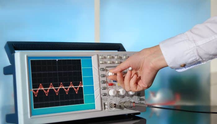
ECoG operates on the fundamental principle of capturing electrical potentials generated by neuronal activity directly from the brain’s surface. To do this, a series of electrodes is placed on the exposed cortex, usually during neurosurgical procedures. These electrodes are much closer to the source of electrical activity than scalp-based EEG electrodes, allowing for a clearer and more detailed signal capture.
The recorded signals primarily reflect the summated activity of large populations of neurons, specifically from the pyramidal cells located in the brain’s cortical layers. Given its direct access, ECoG is less susceptible to the common interferences that affect EEG, such as the scalp, skull, and dura mater.
By analyzing the frequency, amplitude, and patterns of these captured signals, clinicians and researchers can derive a wealth of information about brain function, ranging from the identification of epileptic foci to the intricate details of sensory processing, motor planning, and even cognitive functions.
Equipment and tools used
Electrocorticography requires a combination of specialized equipment to ensure accurate data capture. The primary tool is the ECoG electrode array, which consists of a set of flat disc-shaped electrodes, usually made of platinum or stainless steel. These arrays can vary in size and configuration, ranging from a few individual electrodes to grids containing dozens. Some configurations include:
- Grid Electrodes: These are used for covering larger cortical areas and often have a rectangular arrangement of multiple electrodes.
- Strip Electrodes: Narrow strips used for interhemispheric recordings or for specific sulci.
- Depth Electrodes: These are used for recording from subcortical structures or deeper cortical layers.
Associated with the electrode arrays are the amplifiers, which boost the faint electrical signals from the brain to readable levels, and data acquisition systems that digitize and store the signals. Modern systems often come with software solutions that allow for real-time visualization, analysis, and mapping of brain activity.
Procedure: Placement of electrodes and data acquisition
The placement of ECoG electrodes requires a surgical procedure, which is invasive in nature. Before the procedure:
- Planning: Medical imaging techniques like MRI or CT scans are utilized to plan the exact locations for electrode placement.
- Surgery: The patient undergoes a craniotomy, where a portion of the skull is temporarily removed to expose the brain’s surface. Once the brain is exposed, the electrode array is placed directly onto the cortex. The position of the electrodes is usually verified using intraoperative imaging.
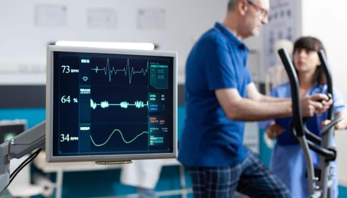
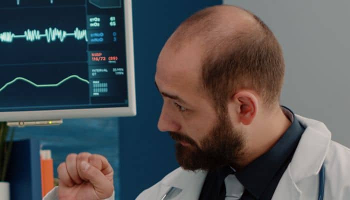
After placement, the electrodes are connected to the amplifiers and data acquisition systems. The brain’s electrical activity is then monitored, either during the surgery itself (for procedures like identifying epileptic foci) or post-operatively (for longer-term monitoring).
The recording period can vary from a few hours to several days, depending on the clinical or research objective. Throughout this period, the patient might be asked to perform specific tasks to elicit particular brain responses.
Once the necessary data is collected, the electrodes are surgically removed, and the craniotomy is closed.
Challenges and Limitations
1. Invasiveness of the procedure: One of the most evident limitations of ECoG is its invasive nature. Unlike non-invasive techniques like EEG, ECoG requires a surgical procedure to place the electrodes directly onto the brain’s surface. This procedure involves inherent risks associated with surgeries, such as potential infections, bleeding, or damage to surrounding brain tissue. Furthermore, the invasiveness of the process limits its applicability mainly to individuals already undergoing brain surgeries, typically for conditions like epilepsy. It’s not a feasible option for routine diagnostics or widespread research applications.
2. Long-term stability of recordings: While ECoG offers a higher resolution and clearer signal than non-invasive techniques, maintaining the stability of these recordings over extended periods can be challenging. The brain’s natural movements, due to activities such as breathing and blood flow, can cause minute shifts in electrode placement, which can influence recording consistency. Moreover, there is the potential for inflammatory reactions over time, which might interfere with the quality of the signals obtained.
3. Interpretation of complex ECoG data: ECoG provides rich data detailing the brain’s electrical activity. However, interpreting this data is not always straightforward. The signals obtained reflect the summation of activities from thousands of neurons, and deciphering specific neuronal actions or connections can be intricate. It requires expertise to differentiate between relevant signals and potential artifacts, to understand the significance of various oscillatory patterns, and to correlate the data with specific brain functions.
4. Scope of Application: While ECoG is invaluable in specific contexts, its application scope can be limited. It excels in pre-surgical mapping and seizure focus identification, but it might not be the tool of choice for other neurological conditions. Additionally, its invasive nature means that it’s often reserved for cases where other non-invasive modalities have been ineffective or inconclusive.
Recent Advances and Future Directions
1. Technological improvements in electrode design: In recent years, there have been significant advancements in electrode design for ECoG. Researchers and engineers have been focusing on developing thinner, more flexible electrodes that can conform better to the brain’s surface. This has the potential to enhance signal quality and reduce tissue irritation. Additionally, innovations in materials science have introduced biocompatible coatings and materials that minimize inflammatory reactions. The integration of micro-electronic systems has also facilitated the creation of high-density electrode arrays, capturing more detailed neural activity and covering broader regions of the brain.
2. Integration with other neuroimaging modalities: One of the promising directions in ECoG research is its integration with other neuroimaging techniques, such as functional MRI (fMRI) or magnetoencephalography (MEG). By combining the high temporal resolution of ECoG with the spatial resolution of techniques like fMRI, researchers can gain a more comprehensive understanding of brain dynamics. This multimodal approach allows for a richer interpretation of neural processes, from the intricate oscillatory patterns seen in ECoG to the blood-oxygen-level-dependent (BOLD) responses in fMRI.
3. Potential applications in neuromodulation therapies: ECoG’s ability to provide real-time, high-resolution data on brain activity is being explored for its potential in neuromodulation therapies. For instance, ECoG could play a role in brain-computer interfaces (BCIs) that assist paralyzed individuals in controlling prosthetic limbs or communication devices. Beyond BCIs, there’s growing interest in using ECoG for real-time feedback in neuromodulation treatments for disorders like depression, Parkinson’s disease, and epilepsy. By monitoring the immediate effects of interventions like deep brain stimulation (DBS) through ECoG, clinicians could adjust treatments for optimal results.