Vascular Doppler ultrasound is a non-invasive diagnostic tool that uses sound waves to evaluate blood flow in the arteries and veins. Learn more about this technique with our comprehensive guide.
Vascular Doppler ultrasound is a medical imaging technique that uses sound waves to evaluate blood flow in the arteries and veins. It is a non-invasive and painless procedure that can provide valuable information about the health of your circulatory system. In this guide, we’ll explore the basics of vascular Doppler ultrasound, including how it works, what to expect during the procedure, and its potential benefits and limitations.
What is Vascular Doppler Ultrasound?
How Does Vascular Doppler Ultrasound Work?
What are the Benefits of Vascular Doppler Ultrasound?
What are the Different Types of Vascular Doppler Ultrasound?
What to Expect During a Vascular Doppler Ultrasound Exam?
1. What is Vascular Doppler Ultrasound?
Vascular Doppler ultrasound is a diagnostic tool that uses high-frequency sound waves to create images of blood flow in the arteries and veins. It is a non-invasive procedure that can help doctors evaluate the health of your circulatory system and diagnose conditions such as blood clots, deep vein thrombosis, and peripheral artery disease. During the procedure, a technician will apply a gel to your skin and use a handheld device called a transducer to send sound waves through your body.
The sound waves bounce off your blood vessels and create images that can be viewed on a computer screen.
2. How Does Vascular Doppler Ultrasound Work?
Vascular Doppler ultrasound works by using high-frequency sound waves to create images of blood flow in the arteries and veins. The sound waves are emitted from a handheld device called a transducer, which is placed on the skin over the area being examined. The sound waves then bounce off the blood cells and tissues in the body, creating echoes that are picked up by the transducer and sent to a computer.
The computer then uses the echoes to create images of the blood flow in real-time, which can be viewed by the technician and the doctor. This allows them to evaluate the health of the circulatory system and diagnose any conditions that may be present.
3. What are the Benefits of Vascular Doppler Ultrasound?
Vascular Doppler ultrasound offers several benefits as a non-invasive diagnostic tool. It does not use radiation, making it a safer option for patients who may be sensitive to radiation or who have already undergone multiple imaging tests.
It is also painless and does not require any injections or incisions, making it a more comfortable option for patients. Additionally, it provides real-time images of blood flow, allowing doctors to quickly diagnose and treat any conditions that may be present. Overall, vascular Doppler ultrasound is a valuable tool in evaluating and monitoring the health of the circulatory system.

4. What are the Different Types of Vascular Doppler Ultrasound?
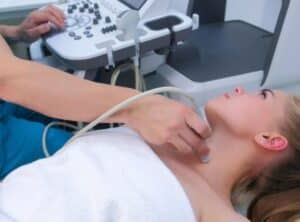
There are several types of vascular Doppler ultrasound, each with its own specific purpose. The most common types include carotid Doppler ultrasound, which evaluates blood flow in the carotid arteries in the neck; venous Doppler ultrasound, which evaluates blood flow in the veins of the legs or arms; and arterial Doppler ultrasound, which evaluates blood flow in the arteries of the legs or arms.
Other types of Doppler ultrasound may be used to evaluate blood flow in specific organs or tissues, such as the liver or kidneys. Your doctor will determine which type of Doppler ultrasound is most appropriate for your specific needs.
5. What to Expect During a Vascular Doppler Ultrasound Exam?
During a vascular Doppler ultrasound exam, you will lie on an examination table while a technician applies a gel to the area being examined. The technician will then use a handheld device called a transducer to send sound waves into the body and record the echoes that bounce back.
These echoes are then converted into images that can be viewed on a monitor. The exam is painless and typically takes between 30 minutes to an hour to complete, depending on the area being examined. After the exam, you can resume your normal activities immediately. Your doctor will review the results of the exam with you and discuss any necessary follow-up care